From 2D to 3D: How Real-Time Imaging Is Transforming Interventional Radiology
Introduction
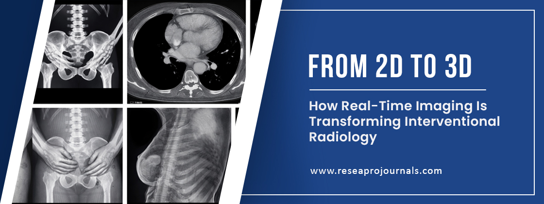
The field of interventional radiology (IR) has undergone a remarkable transformation over the past decade due to rapid advancements in imaging technologies. Traditionally dependent on two-dimensional (2D) fluoroscopy and computed tomography (CT), modern interventional radiology is now increasingly driven by real-time three-dimensional (3D) imaging systems that provide enhanced anatomical visualization, procedural accuracy, and intraoperative guidance.
Real-time imaging has emerged as a critical component in image-guided interventions, enabling clinicians to perform minimally invasive procedures with improved spatial orientation and reduced procedural risk. Technologies such as cone-beam CT (CBCT), augmented fluoroscopy, fusion imaging, intravascular ultrasound (IVUS), and AI-enhanced visualization platforms are fundamentally reshaping diagnostic and therapeutic radiology workflows.
The integration of 3D visualization into interventional procedures is not only improving procedural precision but also contributing to personalized treatment planning, radiation dose optimization, and superior patient outcomes. As healthcare systems increasingly adopt precision medicine frameworks, the evolution from static 2D imaging toward dynamic 3D image-guided intervention represents a major milestone in clinical radiology innovation.
What Is Real-Time 3D Imaging in Interventional Radiology?
Real-time 3D imaging in interventional radiology refers to advanced visualization technologies that generate volumetric anatomical images during minimally invasive procedures. Unlike conventional 2D fluoroscopic systems, which provide flat projection images, 3D imaging systems create spatially accurate anatomical reconstructions that help clinicians visualize complex vascular structures, tumors, organs, and interventional devices in real time.
These imaging modalities combine multiple imaging datasets, including:
- Computed Tomography (CT)
- Magnetic Resonance Imaging (MRI)
- Cone-Beam CT (CBCT)
- Ultrasound Imaging
- Fluoroscopy
- Positron Emission Tomography (PET)
The transition from planar imaging to volumetric navigation significantly improves lesion targeting, catheter navigation, vascular mapping, and device deployment during interventions such as embolization, tumor ablation, thrombectomy, biopsy, and stent placement.
How Does It Work?
Real-time 3D imaging systems operate through the integration of image acquisition hardware, computational reconstruction algorithms, and navigation software platforms.
The workflow generally includes:
1. Image Acquisition
High-resolution imaging data are collected using CT scanners, rotational angiography systems, or MRI platforms. Multiple projection images are captured from various angles.
2. Volumetric Reconstruction
Advanced computational algorithms reconstruct the acquired data into a 3D anatomical model. Machine learning-assisted reconstruction methods further improve image clarity and reduce motion artifacts.
3. Real-Time Visualization
The reconstructed anatomy is displayed dynamically on interventional workstations, allowing physicians to manipulate the viewing angle, zoom, and cross-sectional perspectives during procedures.
4. Instrument Tracking
Navigation software tracks the movement of guidewires, catheters, needles, and robotic devices within the 3D anatomical environment.
5. Image Fusion
Preoperative CT or MRI scans are fused with live fluoroscopic imaging to create hybrid visualization systems that improve procedural targeting accuracy.
Explanation
The clinical significance of real-time 3D imaging lies in its ability to improve procedural confidence and anatomical precision. Conventional 2D fluoroscopy often presents challenges related to depth perception, tissue overlap, and limited spatial orientation. In contrast, 3D visualization technologies enable comprehensive anatomical assessment with improved depth mapping.
For example, in transarterial chemoembolization (TACE) procedures for liver cancer, cone-beam CT allows interventional radiologists to visualize tumor-feeding arteries with significantly greater precision compared to traditional angiography. Similarly, during endovascular aneurysm repair (EVAR), real-time 3D navigation helps optimize stent graft deployment while minimizing vascular complications.
AI-integrated imaging systems are also accelerating workflow automation in interventional radiology. Deep learning algorithms can automatically segment anatomical structures, identify lesions, and assist in trajectory planning during biopsies and ablation therapies.
Furthermore, augmented reality (AR) and mixed reality (MR) technologies are beginning to integrate with interventional imaging systems, providing holographic anatomical visualization during complex procedures. These emerging technologies may redefine the future of image-guided therapy by enabling immersive surgical navigation environments.
Examples of Real-Time Imaging Applications
Tumor Ablation
3D imaging enables precise needle placement during radiofrequency ablation (RFA), microwave ablation, and cryoablation procedures. Accurate tumor localization reduces collateral tissue damage and improves oncological outcomes.
Neurointerventional Procedures
In stroke thrombectomy procedures, real-time 3D angiography assists clinicians in navigating cerebral vasculature rapidly, improving clot retrieval efficiency and reducing neurological injury.
Cardiovascular Interventions
Fusion imaging technologies are extensively used in transcatheter aortic valve replacement (TAVR), cardiac electrophysiology procedures, and peripheral vascular interventions.
Interventional Oncology
Real-time imaging supports targeted drug delivery, embolization therapies, and image-guided biopsies in precision oncology applications.
Robotic-Assisted Interventions
Robotic catheter systems integrated with 3D imaging platforms improve procedural reproducibility, navigation accuracy, and remote intervention capabilities.
Benefits
Enhanced Procedural Accuracy
3D visualization improves anatomical localization and device positioning, minimizing procedural errors.
Reduced Radiation Exposure
Advanced image reconstruction and navigation systems reduce fluoroscopy time and cumulative radiation dose.
Improved Patient Outcomes
Higher targeting precision contributes to reduced complications, shorter hospital stays, and improved therapeutic success rates.
Better Spatial Orientation
Volumetric imaging enables superior depth perception during complex vascular and oncological interventions.
Support for Precision Medicine
Personalized anatomical mapping facilitates patient-specific treatment strategies.
Limitations
High Infrastructure Cost
Implementation of advanced imaging suites requires substantial capital investment and maintenance expenses.
Technical Complexity
Real-time imaging systems demand specialized training and workflow adaptation among radiologists and technologists.
Data Processing Challenges
Large imaging datasets require powerful computational infrastructure and optimized storage systems.
Motion Artifacts
Respiratory and patient movement may affect image quality during live reconstruction.
Limited Accessibility
Many healthcare facilities, particularly in low-resource settings, face barriers to adopting advanced interventional imaging platforms.
Perspectives on the Future of Interventional Radiology
The future of interventional radiology is closely associated with the convergence of artificial intelligence, robotic systems, digital twin technology, and advanced imaging informatics.
Emerging innovations include:
- AI-driven autonomous catheter navigation
- Holographic surgical guidance systems
- Real-time digital anatomy modeling
- Predictive imaging analytics
- Cloud-based image-sharing ecosystems
- 4D imaging with temporal motion tracking
- AI-assisted radiation optimization systems
Researchers are also exploring the application of quantum imaging and photon-counting CT technologies to further improve spatial resolution and tissue characterization. Additionally, the integration of generative AI in radiology reporting may significantly enhance workflow efficiency and clinical decision support.
As minimally invasive medicine continues to expand, real-time imaging technologies will remain central to the development of safer, faster, and more personalized interventional procedures.
The transition from 2D imaging toward real-time 3D visualization represents one of the most significant technological advancements in interventional radiology. By enhancing anatomical accuracy, procedural guidance, and clinical efficiency, modern imaging systems are transforming the way minimally invasive procedures are performed across oncology, cardiology, neurology, and vascular medicine.
The integration of artificial intelligence, fusion imaging, and advanced computational reconstruction is accelerating the evolution of precision-guided intervention. Despite challenges related to cost, infrastructure, and technical complexity, the clinical benefits of real-time 3D imaging continue to drive widespread adoption across healthcare institutions worldwide.
As radiological innovation advances further, the future of interventional radiology will increasingly rely on intelligent, image-guided ecosystems capable of delivering highly personalized and data-driven therapeutic interventions.
If you're working on Surgery and Interventional Radiology, why not take it further? With Reseapro Journals, you can publish faster and reach a global academic audience. Start your submission today and amplify your research impact.
Reviews & Comments
No reviews yet. Be the first to write one.